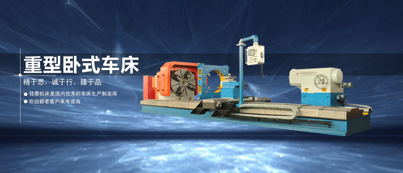
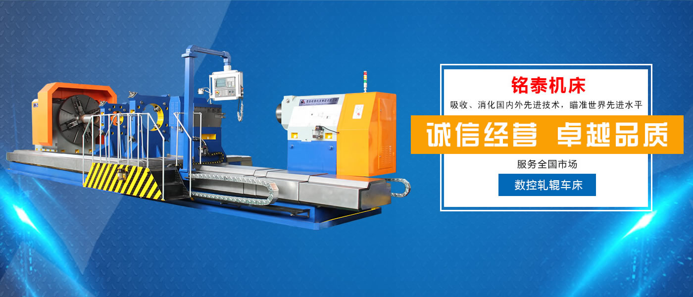
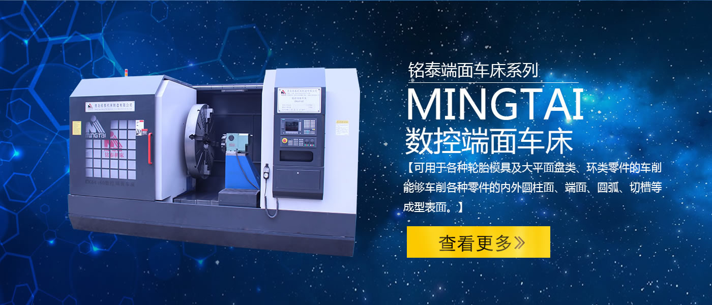
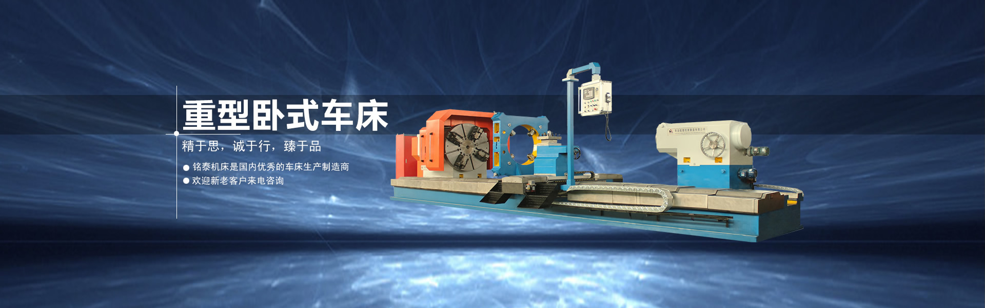
Free Health Care Free Health Care
Main Menu
Bites and Stings
Eye Disorders
General Problems
Infection and Contagious Diseases
Lungs Disorders
AIDS
TETANUS
Hoarseness
Mumps
Nose Bleeds
Skin
Whooping Cough
Amoebiasis
Amenorrhea
Colic Baby
Febrile Seizure

Home :: About Skin

Skin : Structure, Layers & Glands of Skin

The skin consists of three stratified layers-the epidermis, dermis and the subcutaneous tissue from surface downwards. ­

  1. The epidermis consists of several layers of cells.
  2. The corium or dermis consists mostly of connective tissue.
  3. The subcutaneous tissue consists of fatty and fibrous tissue.

The epidermis: The epidermis which consists of many layers of cells is devoid of blood vessels or lymphatics. It gets its nutrition from the underlying dermis which is richly endowed with blood vessels and lymphatics. Epidermis consists of several layers of cells. The stratum corneum which is the outermost layer is composed of dead non-nucleated keratinized cells. Below that, in order are stratum granulosum containing coarse cytoplasmic granules, 5-10 layers of polygonal cells called stratum Malpighii, and the basal cell layer composed of columnar cells. Cells of the basal layer divide progressively and migrate outwards undergoing progressive changes to become keratinized within a period of twenty eight days. The keratinized cells are shed from the surface.

The epidermis contains melanocytes which are derived from the neural crest. The melanocytes produce melanin pigment. They are also found in the matrices of hair follicles. The melanocytes which are dendritic cells derived from neural crest produce melanin from tyrosine through the action of the enzyme tyrosinase. From the melanocytes the melanin passes into the surrounding epidermal cells which passively engulf melanin. Melanogenesis in man is affected by various factors including hormones like ACTH, sex hormones and thyroxine, and physical agents like light and constant irritation.

Other dendritic cells seen in the epidermis are Langerhan's cells which constitute about 4% of the total cell population. They can be identified by staining with gold chloride. These cells are derived from bone marrow. Similar cells are seen in the dermis, lymph­n odes and thymus. They take part in antigen processing and form the afferent limb of the immune response.

Dermo epidermal junction: This is lined by a basement membrane to which the basal cells are attached. This membrane is wavy and dermal papillae project into the epidermis. The dermal papillae are richly supplied with blood vessels and nerves. The nerve fibers ramify between the deeper layers of the epidermis.

Dermis (corium): The dermis is made up of collagenous connective tissue mainly and elastin in smaller amounts interspersed in the ground substance which is made up of mucopolysaccharides. There are also fibroblasts, histiocytes, and mast cells, which are mesenchymal in origin. The dermis shows epidermal down growths of hair follicles, the sebaceous glands and sweat glands. It also contains an extensive network of blood vessels and nerves. The caliber of the dermal blood vessels can be controlled by arteriovenous anastomoses which act as shunts. The blood vessels are under neural and hormonal control as in other parts of the body. This mechanism ensures a proper supply of blood to the cutaneous appendages like hair bulbs, sweat glands and sebaceous glands. The arrangement of dermal blood vessels plays a major role in temperature regulation.

Cutaneous Nerves:- The cutaneous nerves in the dermis are both autonomic and sensory. The arterioles are supplied by adrenergic sympathetic nerves which mediate vasoconstriction. The adrenergic sympathetic fibers also supply arrectores pilorum muscles, and the myoepithelial celIs of the apocrine and eccrine sweat glands. The secretory fibers for the eccrine sweat glands are cholinergic sympathetic. The skin is devoid of any demonstrable vasodilator nerves.

The sensory end-organs conveying pain, temperature, and touch are seen in the dermis. There is no absolute specificity for these receptors except for "Vater-Paccini corpuscles", stimulation of which gives the sensation of pressure. The free nerve endings can convey all modalities of sensation. The sensation of pruritus is mediated by the small unmyelinated nerves which, mainly subserve the sensation of pain. The stimulation of these nerves results in pruritus, the stimuli being less intense than that required to evoke pain.

The Glands of the Skin

a. Sebaceous glands: These are of epidermal origin seen in close proximity of the hair follicIes at the upper part of dermis. They are most numerous on the scalp, face, front of the chest and back and they are absent on the palms and soles. Their secretion, sebum is formed by disintegration of the lining cells and this is not under neural control. The sebum is discharged to the surface through the hair follicle. The sebaceous glands hypertrophy during puberty under hormonal influences.

b. Apocrine glands: They are large coiled, tubular glands opening into the hair follicles and their function is not clearly known. They are seen in the axillae, around the nipples, perineum, and genital regions. Specialized apocrine glands are seen in the eyelids, and in the ear canal. Apocrine glands are surrounded by contractile myoepithelial cells which help to expel their contents. These myoepithelial cells derive their innervation from adrenergic twigs which respond to emotional stimuli.

c. Eccrine glands: They are more numerous over the forehead, axillae, palms, and soles though they are widely distributed throughout the body. The secretory portion is situated in the dermis and this is supplied by cholinergic sympathetic fibers. Adrenergic sympathetic fibers which supply the myoepithelial sheath help to expel their contents. Sweat in the healthy person contains all the electrolytes found in the plasma or interstitial fluid but to a lesser extent. Heat and emotional stress are the main stimuli for sweat secretion 

Home || Feedback ||

(c)Copyright Free-Health-care.com. All rights reserved.

Disclaimer : All information on free-health-care.com is for educational purposes only. For specific medical advice, diagnoses, and treatment, please consult your qualified health care provider. We will not be liable for any complications, or other medical accidents arising from the use of any information on this web site.